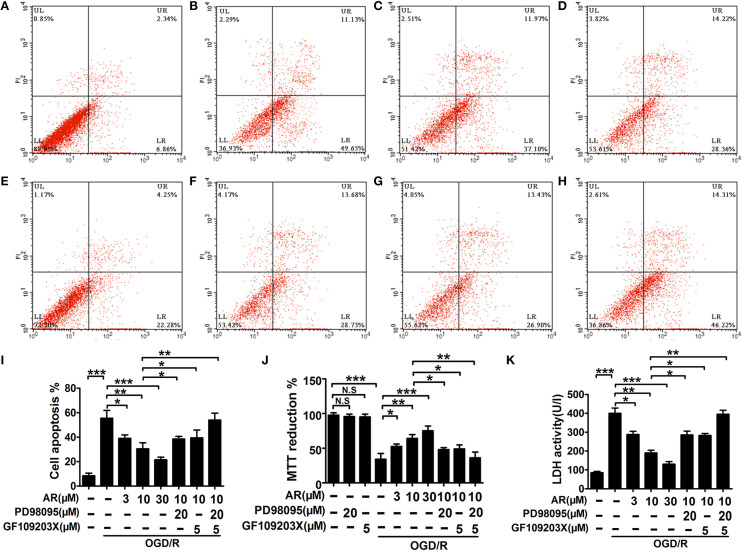
Figure 3.
The effect of AR on apoptosis rates, cell viability, LDH levels in primary cortical neurons. After OGD/R and then followed by 8 h AR (3, 10, and 30 μM) or indicated inhibitors treatment, the OGD/R was applied as illustrated in methods. Apoptosis rates were detected with annexin V/PI double staining after incubation and presented in (A) normal neurons, (B) neurons only experiencing OGD/R, (C) neurons treated with 3 μM AR after OGD/R, (D) neurons treated with 10 μM AR after OGD/R, (E) neurons treated with 30 μM AR after OGD/R, (F) neurons were exposed to PD98095 together with 10 μM AR treatment 8 h after OGD/R, (G) neurons were exposed to GF109203X together with 10 μM AR treatment 8 h after OGD/R, and (H) neurons were exposed to PD98095 and GF109203X together with 10 μM AR treatment 8 h after OGD/R. (I) Quantification of apoptotic rates. (J) Cell viability was analyzed by MTT assay and data were described as % of normal neurons. (K) Neurons injury was determined by LDH activity assay. Results mentioned-above were mean ± SD for 4 individual experiments which, for each condition, were performed in quadruplicate. *P < 0.05; **P < 0.01; ***P < 0.001. N.S, no significant.

