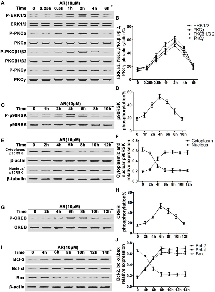
Figure 4.
The effect of AR on the phosphorylation of ERK1/2, cPKC, p90RSK, CREB and bcl-2, bcl-xl and bax expressions in primary cortical neurons after OGD/R. Primary cortical neurons were treated with 10 μM AR for indicated time after OGD/R operation and then protein phosphorylation levels or expressions were determined by western blot assay. (A) Representative result of ERK1/2 and cPKC (PKCα, PKCβ1/β2, PKCγ) phosphorylation alterations in neurons experiencing different time AR treatment, and data from four individual experiments were described in (B). (C) Representative results of p90RSK phosphorylation alterations in neurons experiencing different time AR treatment, and this time-kinetic data were described in (D). (E) Representative result of p90RSK nucleus-cytoplasm distribution alterations in neurons experiencing different time AR treatment, and this time-kinetic data were described in (F). (G) Representative result of CREB phosphorylation alterations in neurons experiencing different time AR treatment, and this time-kinetic data were described in (H). (I) Representative result of bcl-2, bcl-xl, and bax expressions changes in neurons experiencing different time AR treatment, and this time-kinetic data were described in (J). Results mentioned-above were mean ± SD for four individual experiments which, for each condition, were performed in quadruplicate.

