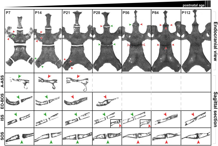
Figure 8.
Postnatal ontogenic changes in the cranial base in C56BL/6J mice. Top row: Endocranial view of representative reconstructed micro-CT scans of P7, P14, P21, P28, P58, P84, and P112 mouse skulls (left to right). Surrounding structures have been digitally cropped for better visualization of the synchondroses of interest. Green arrowheads indicate areas that appear patent while red arrowheads indicate areas where the synchondroses appear closed. Rows two and three shows sagittal sections perpendicular to the transverse width of the ala-alisphenoid (A-ASS), exoccipital-basioccipital (EO-BOS) synchondroses respectively. Rows four and five shows mid-sagittal sections through the intersphenoidal (ISS) and sphenoccipital synchondroses (SOS) respectively. Insets (Row four, P28 and P56) show sagittal sections of the ISS at the lateral end where it appears to be closing (red arrowheads).

