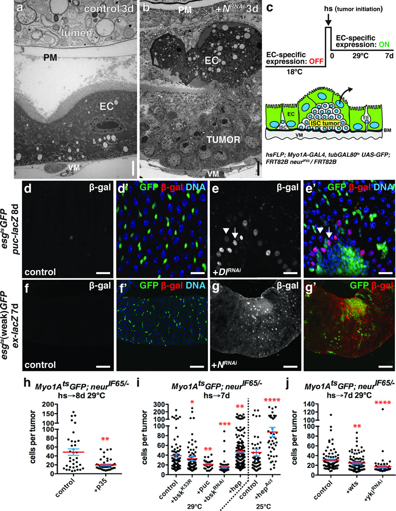
Figure 4. Growing ISC tumors induce changes in the niche.
(a–b) Transmission electron micrograph of anterior midgut expressing GFP (a) or NRNAi (b) with esgts for 3 days.
(c) System to independently initiate ISC- derived tumors by heat shock- induced Flp-FRT mediated recombination and subsequently express transgenes in ECs with Myo1Ats.
(d–e) β-galactosidase (d,e; d’,e’, red) in midguts of flies bearing puc-lacZ and expressing GFP (d’, green) or GFP and DlRNAi (e’, green) with esgts for 8 days. High β-galactosidase (e; e’, red; arrow) was observed in ECs adjacent to ISC tumors; lower β-galactosidase (arrowhead) in ECs further away.
(f–g) β-galactosidase (f,g; f’,g’, red) in midguts of flies bearing ex-lacZ and expressing GFP (f’, green) or GFP and NRNAi (g’, green) with esgts (weak) for 7 days.
(h–j) Cells per neurIF65/− tumor amongst ECs expressing GFP (control, n=35 tumors from 12 midguts, skewness= 1.117, kurtosis= 0.4573) or GFP and p35 (n=42 tumors from 28 midguts, p=0.0019, skewness= 1.586, kurtosis= 1.707) at 29 °C (h); GFP (control, n=83 tumors from 32 midguts, skewness= 2.650, kurtosis= 8.662), GFP and bskK53R (n=82 tumors from 45 midguts, p=0.0346, skewness= 3.805, kurtosis= 17.430), GFP and puc (n=29 tumors from 19 midguts, p=0.0046), GFP and bskRNAi (n=57 tumors from 44 midguts, p<0.0001, skewness= 3.857, kurtosis= 16.530) or GFP and hep (n=191 tumors from 68 midguts, p=0.0045, skewness= 1.508, kurtosis= 2.082) at 29 °C or GFP (control, n=51 tumors from 23 midguts, skewness= 2.414 and kurtosis= 5.719) or GFP and hepAct (n=42 tumors from 14 midguts, p<0.0001, skewness= 1.413 and kurtosis= 1.724) at 25°C (i); GFP (control, n=79 tumors from 41 midguts, skewness= 2.017, kurtosis= 4.577), GFP and wts (n=92 tumors from 33 midguts, p=0.0088) or GFP and ykiRNAi (n=55 tumors from 17 midguts, p<0.0001, skewness= 5.693, kurtosis= 36.85) at 29°C (j) with Myo1Ats, 7–8 days.
In h–j, midguts were pooled from 3 independent experiments; p-values from Mann-Whitney test; mean (red line) and s.e.m. (blue) are shown. DNA in d’, e’, f’ (blue). Scale bars in a–b, 5 µm; in d–d’, 25 µm; in e–e’, 20µm; in f–g’, 60 µm.

