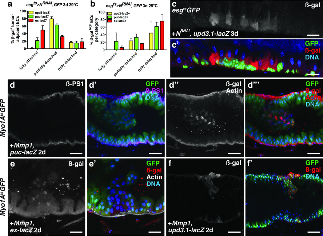
Figure 7. Tumor- induced enterocyte detachment induces JNK and Yki activity and Upd3 expression.
(a) Mean percent with s.e.m. of β-galactosidase (β-gal)- positive N− tumor-adjacent enterocytes that were fully detached, partially detached or fully attached in midguts bearing upd3.1-lacZ (yellow, n=4 z-stacks), puc-lacZ (green, n=3 z-stacks) or ex-lacZ (red, n=3 z-stacks) and expressing GFP and NRNAi with esgts for 3 days.
(b) Mean percent with s.e.m. of N− tumor-adjacent enterocytes in each category (fully detached, partially detached and fully detached) that had high β-galactosidase positivity, in midguts bearing upd3.1-lacZ (yellow, n=4 z-stacks), puc-lacZ (green, n=3 z-stacks) or ex-lacZ (red, n=3 z-stacks) and expressing GFP and NRNAi with esgts for 3 days.
(c) β-galactosidase (red) in midguts bearing upd3.1-lacZ expressing GFP (green) and NRNAi with esgts for 3 days.
(d) β-PS1 integrin (d; d’, magenta) in midguts bearing puc-lacZ and expressing Mmp-1 and GFP (d’, green) with Myo1Ats for 2 days. β-galactosidase (d’’; d’’’, red (nuclear)) in ECs and phalloidin (Actin) (d’’; d’’’, red) in VM of midguts bearing puc-lacZ and expressing Mmp-1 and GFP (d’’’, green) with Myo1Ats for 2 days.
(e) β-galactosidase (e; e’, red) in ECs and phalloidin (e’, white) in VM of midguts bearing ex-lacZ and expressing Mmp1 and GFP (e’, green) with Myo1Ats for 2 days.
f) β-galactosidase (f; f’, red) in ECs of midguts bearing upd3.1-lacZ and expressing Mmp1 and GFP (f’, green) with Myo1Ats for 2 days.
In a–b, z-stacks acquired from 2 independent experiments. DNA in c’, d’, d’’’, e’ and f’ (blue). Scale bars in c–c’, 40µm; d–d’’’, 35µm; e–e’, 25 µm; f–f’, 60µm.

