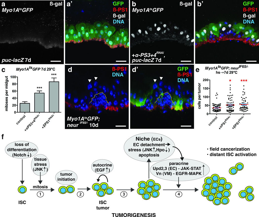
Figure 8. Integrin loss from enterocytes induces JNK activity and promotes ISC tumor growth.
(a–b) β-galactosidase (nuclear) (a,b ; a’,b’, white) and β-PS1 (a’, b’, red) in ECs of midguts bearing puc-lacZ and expressing GFP (a’, green) or GFP and αPS3+4RNAi (b’, green) with Myo1Ats for 7 days.
(c) Mean number with s.e.m. of phosphorylated histone H3 Ser10 positive cells per midgut in flies expressing GFP (control, n=18 midguts), GFP and αPS3+4RNAi (n=19 midguts, Mann-Whitney test: p<0.0006) or βPS1RNAi (n=17 midguts, Mann-Whitney: p<0.0004) with Myo1Ats for 7 days.
(d) β-PS1 integrin (d, d’, red) in midguts bearing neurIF65/− tumors (yellow dashed line) and expressing GFP (d’, green) with Myo1Ats for 10 days. Arrowheads indicate detached ECs apical to tumors that have lost basal β-PS1 expression.
(e) Cells per neurIF65/− tumor with mean (red line) and s.e.m. amongst ECs expressing either GFP (control, n=74 tumors from 35 midguts, skewness= 1.876, kurtosis= 4.283) or GFP and αPS1RNAi (n=63 tumors from 10 midguts, Mann-Whitney: p= 0.0116, skewness= 1.221, kurtosis= 1.126) or GFP and βPS1RNAi (n=54 tumors from 23 midguts, Mann-Whitney: p= 0.0004, skewness= 1.262, kurtosis= 1.093) with Myo1Ats for 7 days.
(f) Model for Notch-dependent tumorigenesis in the adult Drosophila midgut.
In c and e, midguts pooled from 3 independent experiments. DNA in a’, b’, d–d’ (blue). The scale bars in a-b’ and d–d’, 20 µm.

