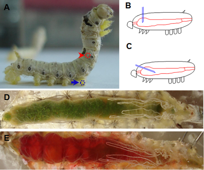
Figure 1. Insect midgut puncture and injection.

(A) Position for making a needle puncture (arrowhead-indicated) in the midgut of a silkworm larva after the corresponding skin was sterilized. A control wound was made in one of the hind-legs as indicated by the arrow. (B,C) A diagram to show how needle puncture (B) and injection (C) were performed. A puncture was made by vertically probing the integument with half of the needle inside the body (B). If a solution was injected into the midgut, the needle was turned towards the hind of midgut (C). Morphology of a naïve midgut (D) or a midgut injected with 50 μl neutral red after 30 min (E). Less neutral red leaked with the hemocoel.
