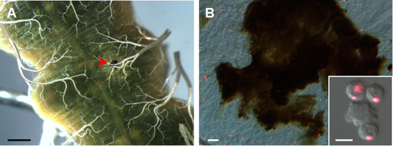
Figure 2. Circulating hemocytes are involved in midgut wound repair.
(A) A melanized scab produced over the wound site. At 6 h after the wound was made, the midgut was dissected to show a melanized scab (arrowhead). (B) Hemocytes in the scab. Circulating hemocytes were pre-labeled via phagocytosis of injected fluorescent beads for at least 6 h as described25 and then a needle puncture was made. The inset is a picture to show hemocytes that had phagocytosed red fluorescent beads. The melanized scab was removed and pressed on a slide to observe fluorescent beads. The pictures were merged from those taken using a red filter and DIC optics. Bar: A, 1 mm; B, 20 μm; Inset in (B), 10 μm.

