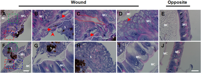
Figure 4. Close observation of the midgut wounds.
At 6 h, the wound was undergoing repair (A–D). At 48 h, the wound was repaired as evidenced by the intact structure (F–I). The part of the midgut opposite the wound site at each time point is presented (E,J). At 6 h (A), the areas framed in red (B), blue (C) and green (D) lines were magnified for closer observation, respectively. At 48 h (F), the areas framed in red (G), blue (H) and green (I) lines were also magnified and the midgut and aggregated hemocytes in the scabs were compared. In (B,C), the arrows point to large muscles. The arrowhead points to a hemocyte connecting a muscle and the scab. The red line framed area in (C) indicates the puncture hole in the midgut. In (C), the arrowhead points to a bubble-like material. All white dotted lines separate the scab and midgut. MG, midgut; S, scab; MC, midgut contents. Bar: (A,F), 100 μm; (B,C,D,G,H,I), 25 μm.

