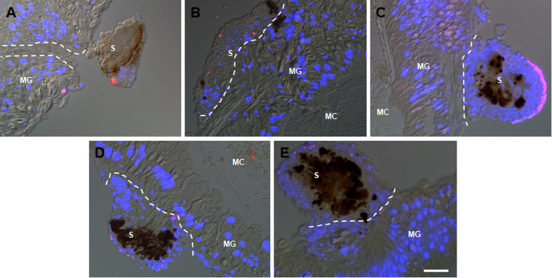
Figure 5. The puncture made in the midgut does not induce apoptosis in cells surrounding the wound.
At 1 (A), 3 (B), 6 (C), 12 (D), and 24 h (E) after the punctures were made, the punctured midguts were dissected for the detection of apoptotic cells using the TUNEL method. Few midgut cells with apoptotic signals were detected around the wound. Some cells with apoptotic signals (red spots) were found in the scab. The white dotted lines separate the scabs and midguts. The pictures were merged from those taken using a red filter (TUNEL), blue filter (DAPI) and DIC optics. MG, midgut; S, scab; MC, midgut contents. Bar: 50 μm.

