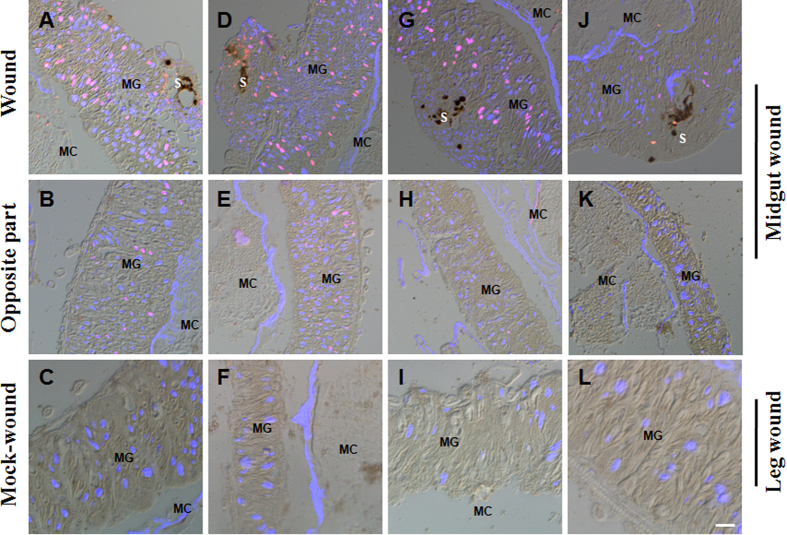
Figure 6. DNA duplication in cells surrounding the wound site.
Silkworm larvae (48 h post ecdysis; day 3 of 5th feeding stage) that received wounds in the midguts or hind-legs were injected with BrdU to label DNA-duplicated cells at 3 (A–C), 6 (D–F), 24 (G–I), and 48 h (J–L), respectively. After the wounds were made, DNA duplication was assayed in cells surrounding the midgut wound site (A,D,G,J), the section opposite the midgut wound (B,E,H,K) and the region corresponding to the area above the midgut wound when punctures were made in one hind-leg (C,F,I,L). Many cells around the wound site had incorporated BrdU (red) according to the staining results. Some cells opposite the wound also incorporated BrdU. The wound in the hind-leg did not induce DNA duplication in the midgut cells. The pictures were merged from those taken using a red filter (BrdU), blue filter (DAPI) and DIC optics. MG, midgut; S, scab; MC, midgut contents. Bar: 50 μm.

