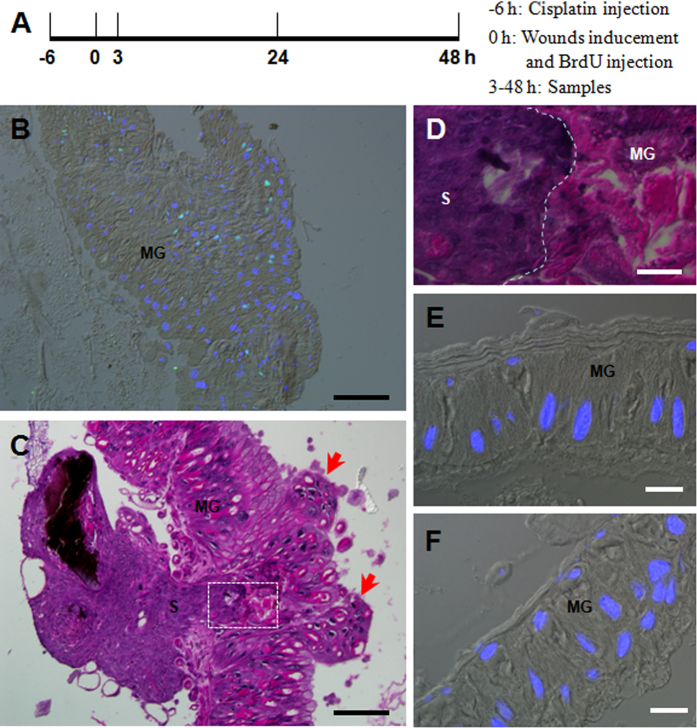
Figure 7. DNA duplication is essential to needle-puncture wound repair in the midguts.
(A) A diagram to show cisplatin injection (-6 h), wound inducement and BrdU injection (0 h) in order to understand the importance of DNA duplication to the wound repair process. Samples were taken at 3 h to detect BrdU incorporation, and at 24 and 48 h for hematoxylin and eosin staining. (B) DNA duplication in cells surrounding the wound at 3 h since it was made (i.e., 9 h after cisplatin injection). The picture was merged from those taken using a red filter (BrdU), blue filter (DAPI) and DIC optics. (C) Morphology of the wound at 48 h (i.e., 54 h after cisplatin injection). The wound was not repaired and many cells were sloughed off the midgut (arrow-indicated). (D) A close-up observation of the neighboring sites of scab (S) and midgut (MG) as outlined in white in (C). The white dotted line indicates the neighboring surface between the hemocyte-aggregated plug-like scab and the midgut. (E,F) Injection of cisplatin did not induce cell apoptosis in midgut cells of naive larvae (E) or larvae that had received a needle-puncture 48 h previously (F). In (F), the injection area was outside of the needle-puncture wound region. The pictures were merged from those taken using a blue filter (DAPI) and DIC optics. Bar: (B,C), 100 μm; (D–F), 20 μm.

