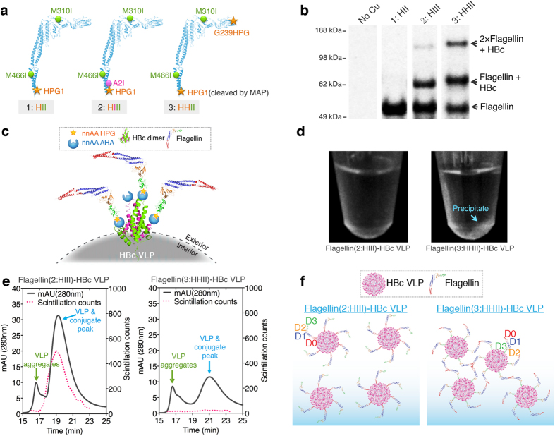
Figure 5. Selection of nnAA attachment sites in flagellin.
(a) Three-dimensional structures of flagellin showing the mutation positions of three flagellin mutants (HII, HPG1+M310I+M466I; HIII, HPG1+A2I+M310I+M466I; and HHII, HPG1+G239HPG+M310I+M466I). (b) Reducing SDS-PAGE autoradiogram analysis of the click reaction products after attaching flagellin mutants to HBc VLPs. The molar ratio of flagellin (52.7 kDa) to HBc monomer (16.7 kDa) in the reactions was 1:2. (c) Diagram of HHII flagellins conjugated to HBc VLPs. There are two nnAA AHAs in each HBc monomer: at the N-terminus and at the 76 site. (d) Photographs showing precipitation of the click reaction products after HHII attachment to the VLP. (e) Size-exclusion HPLC profiles of the click-reaction supernatants with radioactive flagellin and non-radioactive HBc VLPs. (f) Diagrams suggesting a probable cause for the HHII flagellin-HBc conjugate precipitation.

