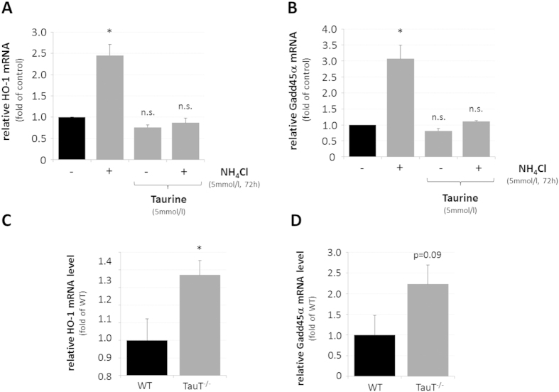
Figure 5. Effect of taurine on HO-1 and Gadd45α mRNA levels in ammonia-exposed cultured astrocytes and in mouse cerebral cortex.
Cultured astrocytes were exposed to NH4Cl (5 mmol/l) or were left untreated (control) for 72 h before RNA was isolated and analysed for HO-1 (A) or Gadd45α (B) mRNA levels by realtime-PCR. Where indicated, astrocytes were treated with taurine (5 mmol/l, 16 h pretreatment). Quantification of (C) HO-1 or (D) Gadd45α mRNA levels by realtime-PCR in wildtype (WT) and taurine transporter knockout (TauT−/−) mice. *statistically significantly different compared to the respected control. n.s.: not statistically significantly different. Data are from 3–10 independent experiments.

