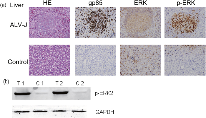
Figure 13. Immunohistochemical and western blot analysis of tumor cells from the liver of ALV-J-infected chickens.
(a) Fresh tissues were collected and fixed in 10% neutralized buffered formalin, dehydrated, embedded in paraffin wax, and then sliced into 6-μm sections. These sections were routinely stained with hematoxylin and eosin (HE), and examined microscopically. Tissues were further immunohistochemically labeled for gp85, ERK or p-ERK with JE9 monoclonal, anti-p44/42 MAPK or anti-phospho-p44/42 MAPK (Thr202/Tyr204) antibodies, respectively. Bar = 100 μm. (b) Western blot analysis detected high expression of p-ERK proteins in myeloid leukosis (ML) samples, but not in the normal samples. Sample identification numbers are indicated above the panel as follows: T, tumor samples; C, non-tumor samples.

