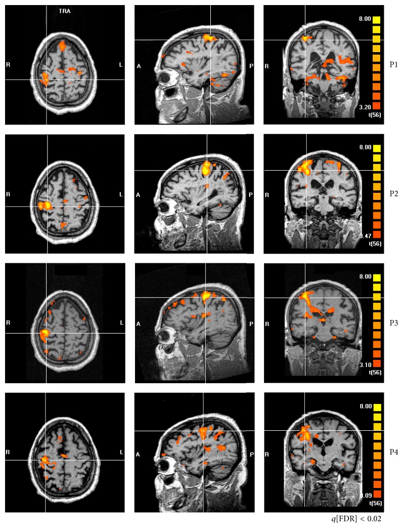
Figure 2.
fMRI of a representative patient (#12) in all periods of evaluation (P1, P2, P3, and P4). Cross-lines are centered over M1 of the ipsilesional hemisphere. At P1, bilateral M1 activity, asymmetrical to the ipsilesional hemisphere. The maps are very sparse, particularly at P1. At P2, sparseness is reduced, and the activity is more confined to M1 and SMA. One month without rehabilitation (P3), the patterns become somehow similar to what they were before treatment onset, which is maintained three months after the end of treatment (P4).

