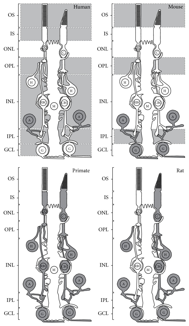
Figure 2.

Schematic illustration representing the distribution of CB1R in the adult retina of several species. CB1R expression was demonstrated in dark gray retinal cells, while CB1R presence was noted in light gray retinal layers without precise localization. OS, outer segments of photoreceptors; IS, inner segments of photoreceptors; ONL, outer nuclear layer; OPL, outer plexiform layer; INL, inner nuclear layer; IPL, inner plexiform layer; GCL, ganglion cell layer; C, cones; R, rods; H, horizontal cells; CBC, cone bipolar cells; RBC, rod bipolar cells; A, amacrine cells; G, ganglion cells; M, Müller cells.
