
Figure 2.
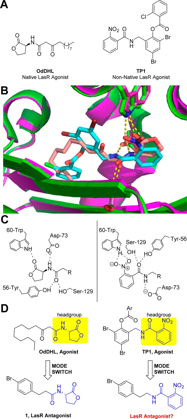
(A) Structures of the native LasR agonist OdDHL and non-native LasR agonist TP1. (B) Overlay of the ligand-binding sites in the OdDHL:LasR (pink) and TP1:LasR (green) X-ray crystal structures with many of the key hydrogen bonds indicated (dashed yellow lines). OdDHL is peach, and TP1 is cyan. Structures are from PDB IDs 3IX3 and 3IX4, respectively. (C) Two-dimensional view of the key hydrogen bonds between LasR and agonists OdDHL and TP1. (D) Illustration of our scaffold-hopping mode-switching approach, installing a p-bromo phenethyl amine on the TP1 headgroup to mimic known AHL-derived LasR antagonist 1.