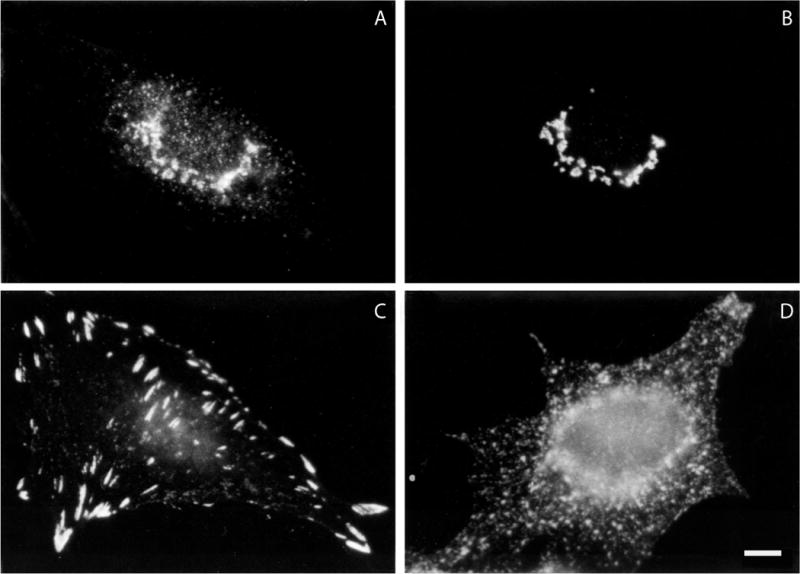
Figure 4.3.1.

Examples of immunofluorescence labeling of formaldehyde-fixed cells. (A) and (B) Double labeling of a normal rat kidney cell with a mouse monoclonal antibody to (A) the β-COP component of coatomer and (B) a rabbit polyclonal antibody to mannosidase II. (C) Distribution of vinculin in a formaldehyde-fixed normal rat kidney cell using a mouse monoclonal antibody. (D) Distribution of transferrin receptor in formaldehyde-fixed HeLa cells using a mouse monoclonal antibody. Bar is equal to 10 μm.
