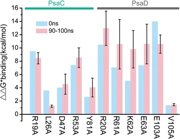
Fig. 4.

Binding affinities estimated from computational alanine scanning for mutations of the OsPsaC-OsPsaD complex ((ΔΔG ∗binding = ΔG ∗binding(mutant) − ΔG ∗binding(wild ‐ type)). ΔΔG ∗binding results for 11 single mutations in either OsPsaC or OsPsaD in the initial structure at 0 ns (green) and averaged over 10 structures at the end of the 100 ns simulation (pink; error bars represent standard deviations)
