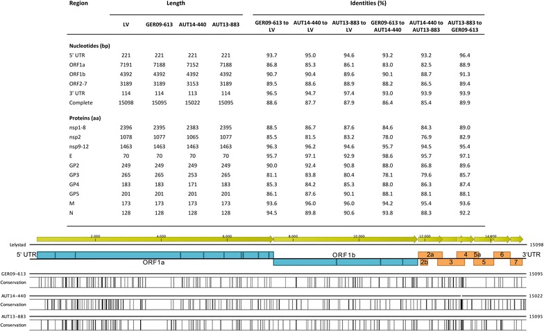
Figure 2.

Detailed comparison of complete genomes of GER09-613, AUT14-440, AUT13-883 and Lelystad virus (LV) (table) and schematic overview of nucleotide differences between LV and the isolates presented in this study. Black bars represent areas with a low degree of similarity to LV. Open reading frames (ORF) of PRRSV that encode non-structural proteins (blue) and structural proteins (orange) are shown.
