
Fig. 2.
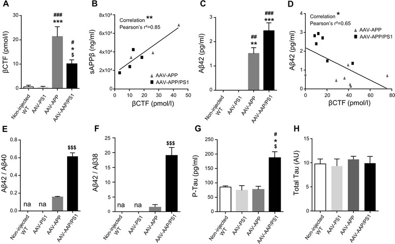
Exogenous human APP is processed following the amyloidogenic pathway, 3 months after injection. C57Bl/6 J mice (all males) were injected at 8 weeks of age either with AAV-CAG-PS1M146L (AAV-PS1 mice), AAV-CAG-APPSL (AAV-APP mice) or both vectors at the same doses as for the other two groups (AAV-APP/PS1 mice, n = 7-8 mice per group). Non-injected WT mice (n = 4) were also analyzed. Mice were killed three months later for analyses of whole hippocampi. a Comparative analysis of TBS-Tx soluble human βCTF levels by ELISA. Note that βCTF levels follow the same pattern of expression than for the human APP in the four different groups (see Fig. 1e). Bars represent means ± SEM. Statistical analysis was performed by one-way ANOVA with Tukey’s post-hoc test where ###, *** denote p < 0.001 versus non-injected WT and AAV-PS1 mice. #, * and $ denote p < 0.05. b Correlation between TBS-Tx soluble human βCTF and sAPPβ levels between AAV-APP and AAV-APP/PS1 mice (n = 7). Linear regression analysis confirms the engagement in the amyloidogenic pathway. Correlation analysis was performed with Pearson’s parametric correlation test: **p < 0.01. c Comparative analysis of TBS-Tx soluble human Aβ42 levels by MSD immunoassay showing higher levels in AAV-APP/PS1 mice (n = 6-8 mice per group). Bars indicate means ± SEM. Statistical analysis was performed by one-way ANOVA followed by Tukey’s post-hoc test where ###, *** denote p < 0.001 versus non-injected WT mice and AAV-PS1 mice. ## and ** denote p < 0.01. d Correlation between TBS-Tx soluble human Aβ42 and βCTF levels between AAV-APP and AAV-APP/PS1 (n = 13). Correlation analysis was performed with Pearson’s parametric correlation test: *p < 0.05. e-f Representation of Aβ42/40 (e) and Aβ42/38 (f) ratios determined by multiplex MSD immunoassay (n = 4 mice per group). Bars indicate means ± SEM. Statistical analysis was performed by one-way ANOVA followed by Tukey’s post-hoc test where $$$ denotes p < 0.001 versus AAV-APP mice. na = not applicable. g Comparative analysis of TBS-Tx soluble murine phosphorylated Tau protein (AT270, Thr181) by ELISA. Bars indicate means ± SEM. Statistical analysis was performed by one-way ANOVA followed by Tukey’s post-hoc test where #, * and $ denote p < 0.05 versus non-injected WT mice, AAV-PS1 mice and AAV-APP mice respectively. h Densitometric analyses of western blots showing the expression of the total Tau protein in each group (n = 4 mice per group). Bars represent means ± SEM and data were normalized with respect to GAPDH