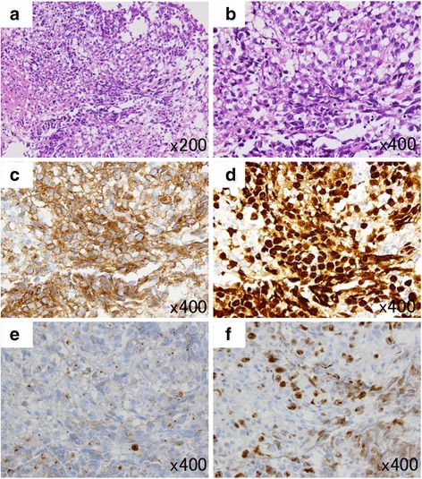
Fig. 2.

Histological findings of the tumor a and b; h-e stain of the tumor. The tumor is composed of sheets and lobules of large and pale cells with often indistinct cell membranes and somewhat vacuolated cytoplasm. Locally, lymphocytic infiltration is present. c-f Immunohistochemically, tumor cells are positive for c-Kit (c) and strongly positive for SALL4 (d). Also, tumor cells are weakly positive for PLAP (e). The Ki-67 labeling index is high in the tumor cells (f). (Original magnification; A × 200, B-F × 400)
