
Fig. 1.
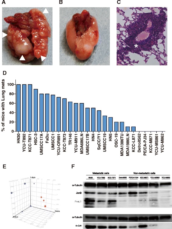
The distant metastatic potential of 26 HNSCC lines in the experimental lung metastatic mouse model. a Lung metastasis in the experimental lung metastatic mouse model of HNSCC. (△ metastatic lesion). b No metastasis was observed in the lungs of the mouse model. c H&E staining of lung metastasis in the experimental lung metastatic mouse model of HNSCC (* metastatic lesion). d Incidence of lung metastasis for 26 cell lines in the experimental lung metastatic mouse model of HNSCC. Three HNSCC cell lines (HN30, KCC-T871, YCU-T892) established 100 % lung metastases, while 5 cell lines (Detroit562, PE/CA-PJ34, KCC-M871, YCU-MS861, YCU-M862) did not establish lung metastasis in any of the mice injected. e The results of principle component analysis (PCA). PCA was performed based on the expression profiles of samples. The first 3 PCAs for 8 HNSCC cells were plotted.  : Metastatic lines (HN30, KCC-T871, YCU-T892),
  : Non-metastatic lines (Detroit562, PE/CA-PJ34, KCC-M871, YCU-MS861, YCU-M862). f Expression of AP-1 family proteins in HNSCC cells by Western blotting. The lung metastatic HNSCC cell lines (HN30, YCU-T892 and KCC-T871) showed higher levels of c-Jun, FosL1 and JunB expression than did the non-metastatic HNSCC cell lines. α-Tubulin was used as an internal control