Fig. 2.

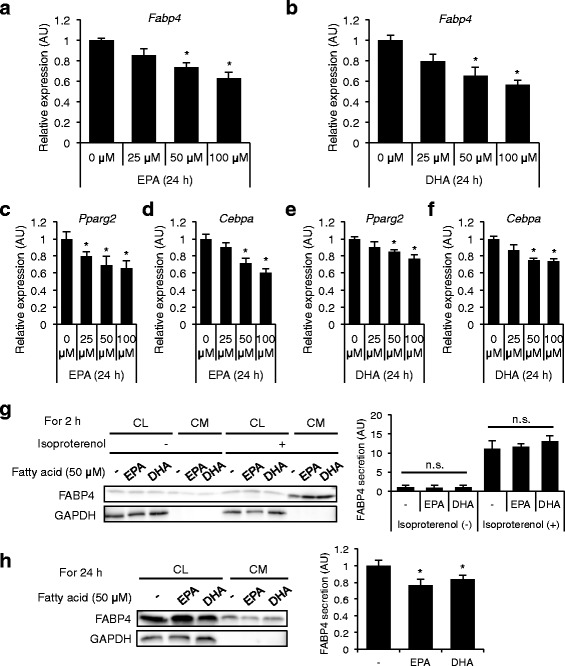
Gene expression and secretion of FABP4 in 3T3-L1 adipocytes treated with omega-3 fatty acids (Study 2). a, b. Gene expression of FABP4 was determined by quantitative real-time PCR in differentiated 3T3-L1 adipocytes treated with 0–100 μM eicosapentaenoic acid (EPA) (A) or 0–100 μM docosahexaenoic acid (DHA) (B) for 24 h (n = 3 in each group). *P < 0.05 vs. 0 μM. c-f. Gene expression of peroxisome proliferator-activated receptor γ2 (PPARγ2) and CCAAT/enhancer binding protein α (C/EBPα) was determined by quantitative real-time PCR in differentiated 3T3-L1 adipocytes treated with 0–100 μM EPA (c, d) or 0–100 μM DHA (E, F) for 24 h (n = 6 in each group). *P < 0.05 vs. 0 μM. g. Western blot analysis of FABP4 and glyceraldehyde 3-phosphate dehydrogenase (GAPDH) using the cell lysate (CL) and conditioned medium (CM) of 3T3-L1 adipocytes treated with 50 μM EPA or 50 μM DHA in the absence and presence of 10 μM isoproterenol for 2 h (n = 5 in each group). n.s., not significant. h. Western blot analysis of FABP4 and GAPDH using the CL and CM of 3T3-L1 adipocytes treated with 50 μM EPA or 50 μM DHA for 24 h (n = 5 in each group). FABP4 secretion was relatively expressed as densitometry of FABP4 in the CM divided by those of FABP4 in the CL and GAPDH in the CL. AU, arbitrary unit. *P < 0.05 vs. Fatty acid (−)
