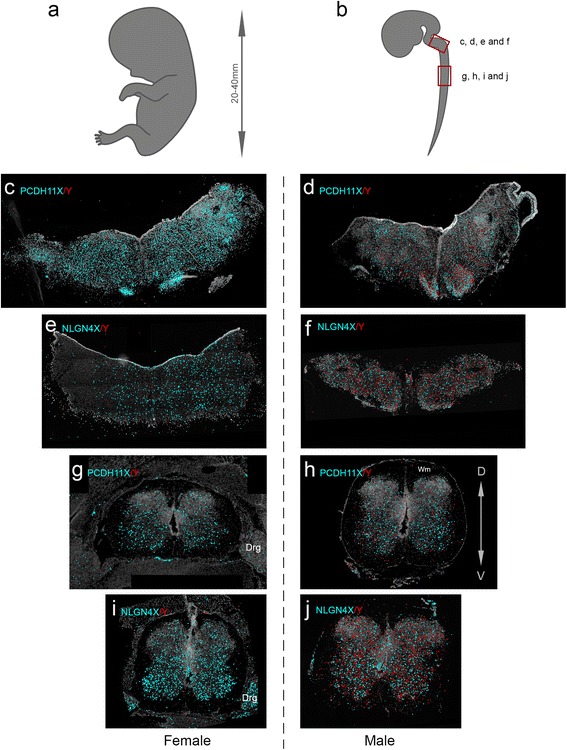
Fig. 2.

Padlock probe hybridization can distinguish between expression of X and Y homolog genes for PCDH11 and NLGN4. a, b Schematic representation of an embryo modified from Gasser [47] and the CNS of an embryo at 12 weeks of gestation modified from His [48]. The boxes in red mark the approximate position of the coronal sections shown in the rest of the figure. c Female medulla oblongata (MO) section hybridized with padlock probes for PCDH11X and Y. X signals in cyan and Y signals in red for all subfigures. d Male MO section hybridized with padlock probes for PCDH11X and Y. e Female MO section hybridized with padlock probes for NLGNX and Y. f Male MO section hybridized with padlock probes for NLGNX and Y. g Female spinal cord (SC) section hybridized with padlock probes PCDH11X and Y. h Male SC section hybridized with padlock probes for PCDH11X and Y. i Female SC section hybridized with padlock probes NLGNX and Y. j Male SC section hybridized with padlock probes for NLGNX and Y. All signals from females and males were detected using a Zeiss Axio Imager.Z2 epi-fluorescence microscope. The images were produced using the Zen software and enhanced to 15–20-pixel dots to allow visualization in ×20 magnification pictures. Drg dorsal root ganglia
