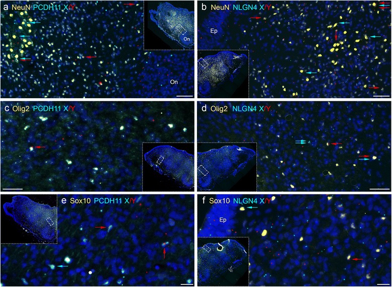
Fig. 7.

Combined padlock hybridization and immunohistochemistry for PCDH11X/Y and NLGNX/Y in MO samples from human male embryos. a, b Padlock probe hybridizations with probes for PCDH11X and PCDH11Y (a) or NLGN4X and NLGN4Y (b) were combined with immunohistochemistry using NeuN antibodies. NeuN-positive cells are stained in yellow, PCDH11X signals in sky blue and PCDH11Y in red. DAPI staining in dark blue allows visualization of nuclei. Each subfigure is an enlargement of the MO tissue showed in each inset picture. Some NeuN+ cells contain signals for PCDH11X (marked with blue arrows), PCDH11Y (red arrows) or both X and Y transcripts (blue and red arrows). c, d Padlock probe hybridization with probes for PCDH11X/Y (a) or NLGN4X/Y (b) was combined with immunohistochemistry using Olig2 antibodies. d, f Padlock probe hybridization with probes for PCDH11X/Y (a) or NLGN4X/Y (b) was combined with immunohistochemistry using Sox10 antibodies. Ep ependymal layer
