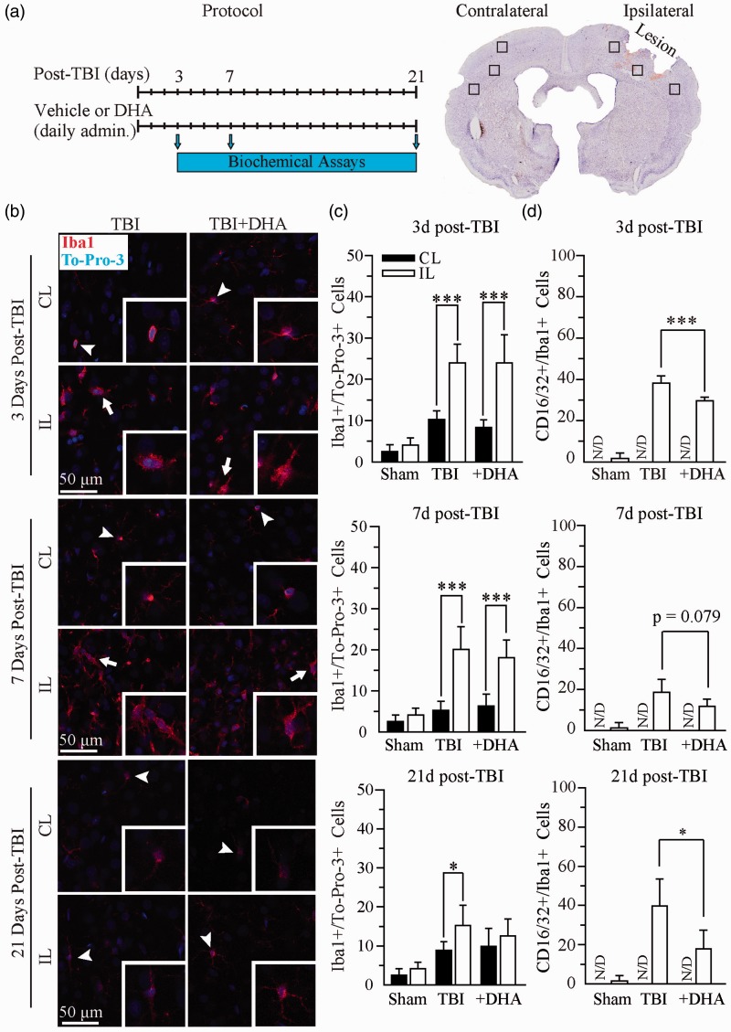
Figure 1.
Iba1+ microglial or macrophage population was not reduced in the DHA-treated TBI brains. (a) Experimental protocol and location of data collection. (b) Confocal images of Iba1+ microglial or macrophage in the IL and CL frontal cortices at 3, 7, and 21 days post-TBI. (c) Summary data of Iba1+ cells/TO-PRO-3+ cells. Values are mean ± SE (n = 4). (d) Summary data of CD16/32+/Iba1+ immunopositive cells. Values are mean ± SE (n = 4). *p < .05, **p < .01, ***p < .001, N/D = no data, one-way ANOVA tests used.

