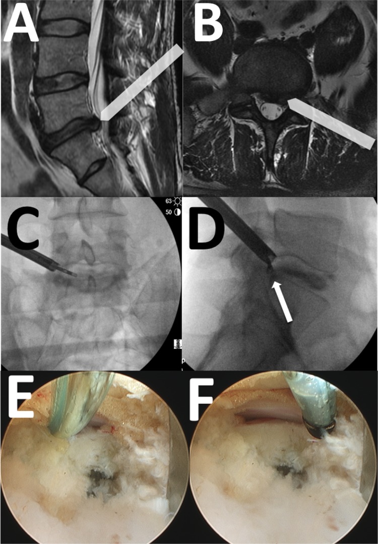
Fig. 1.
A) Sagittal T2 image of the lumbar spine showing an L5-S1 disk herniation. The arrow-bar shows the trajectory of the endoscopic cannula in the cephalad-caudal direction. B) T2 Axial image of the L5-S1 level showing a left-sided disk herniation. The arrow-bar shows the trajectory of the endoscopic cannula in the lateral-medial direction. C) Antero-posterior c-arm image showing the curved grasper reaching toward the disk herniation. D) Lateral c-arm image of the same patient showing the grasper at the posterior disk margin, corresponding to the location of the disk herniation (white arrow). E) Intraoperative photograph with the flexible curved probe (Trigger-flex, Elliquence, Baldwin, New York) inserted between the traversing nerve and the posterior annulus. F) Intraoperative photograph of the Ellman probe pulled back, showing the lateral edge of the traversing nerve root/dural tube.

