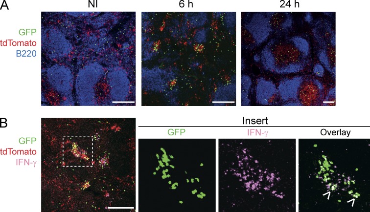
Figure 6.
RP-associated XCR1+ DCs form clusters around the marginal zone and activate mCTLs. Memory Karma mice were generated as described in Fig. 3. Anti-GFP (green) and anti-dsRed (red) antibodies were used to detect memory OT-I cells and tdTomato+ XCR1+ DCs, respectively, on spleen sections imaged by confocal microscopy. (A) Spleen sections were stained with anti-B220 (dark blue) to define B cell follicles. (B) IFN-γ staining (pink) of spleen sections 6 h after secondary Lm-OVA infection. Overlay of IFN-γ staining with GFP gave a white color (arrows in insert). One representative experiment out of three with three mice per group is shown. Bars, 200 µm.

