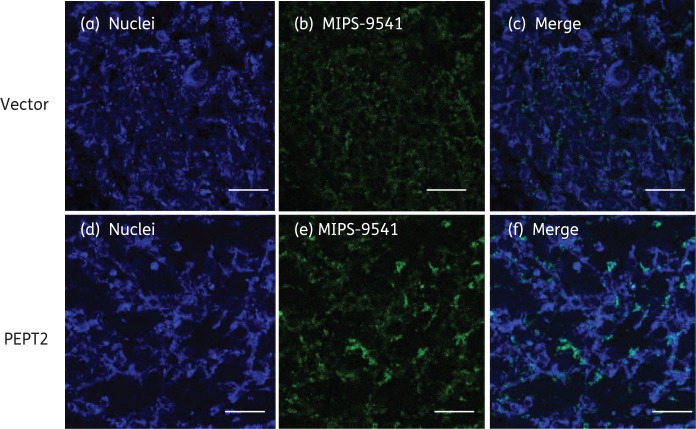
Figure 5.
Fluorescence imaging of MIPS-9541 accumulation in HEK293 cells transfected with PEPT2 or the vector alone. MIPS-9541 (10 µM) was applied to HEK293 cells overexpressing PEPT2 or the vector for 5 min at 37°C. Cells were washed with cold PBS (pH 7.4) three times and mounted in SlowFade® Gold Antifade Mountant with DAPI. Panels (a) and (d) show nuclei staining (blue), panels (b) and (e) show MIPS-9541 accumulated in cells (green), panel (c) shows the merged images of panels (a) and (b) and panel (f) shows the merged images of panels (d) and (e). Scale bars = 50 µm.

