
FIGURE 4:
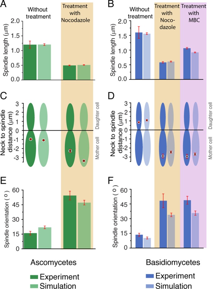
Comparison of in vivo and in silico results upon altering dynamics of MTs in ascomycetes and basidiomycetes. (A and B) Metaphase spindle lengths upon treatment with nocodazole or MBC are plotted for ascomycetes and basidiomycetes along with the unperturbed (dimethyl sulfoxide [DMSO] control) numerical and experimental data. For ascomycetes, we observed that, upon nocodazole treatment, the spindle length becomes ∼0.50 ± 0.04 μm, which is in accordance with our model prediction ∼0.52 ± 0.01 μm. Similarly, in basidiomycetes, the spindle length is reduced to ∼0.61 μm from its wild-type spindle length of ∼1.66 μm. This is in accordance with the experimental value 0.62 ± 0.04 μm. For MBC treatment, in basidiomycetes, the spindle length is shortened to 0.9 μm from its native value of 1.6 μm. This in silico result is in agreement with the experimental data for basidiomycetes as shown. (C and D) Measurements of spindle to neck distances for ascomycetes (with nocodazole) and basidiomycetes (either with nocodazole or MBC) revealed the inability of the spindles to move to their unperturbed spatial locations. The spindle always remained in the mother cell with an increased mean distance (∼ −2.7 μm for nocodazole) from the neck compared with its wild-type value (∼ −1.0 μm) in ascomycetes. In basidiomycetes, the spindle failed to move to the bud, and always remained in the mother cell during either nocodazole or MBC treatment. (E and F) The spindle orientation in the presence or absence of drugs was measured, and it was found to misalign with mother–daughter cell axis, as shown by the higher spindle orientation angle in both cases. Red bars indicate SEM.