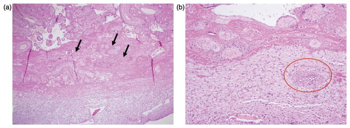
Figure 1.
Histopathological examination of the placenta from a previous pregnancy showed fibrinoid deposition (arrow) in the intervillous space surrounding more than 50% of the villi in some full-thickness sections (H&E; 40×) (a) and absence of physiologic transformation of a spiral artery, i.e. persistent muscularization (circle) in the basal plate (H&E, 100×) (b).

