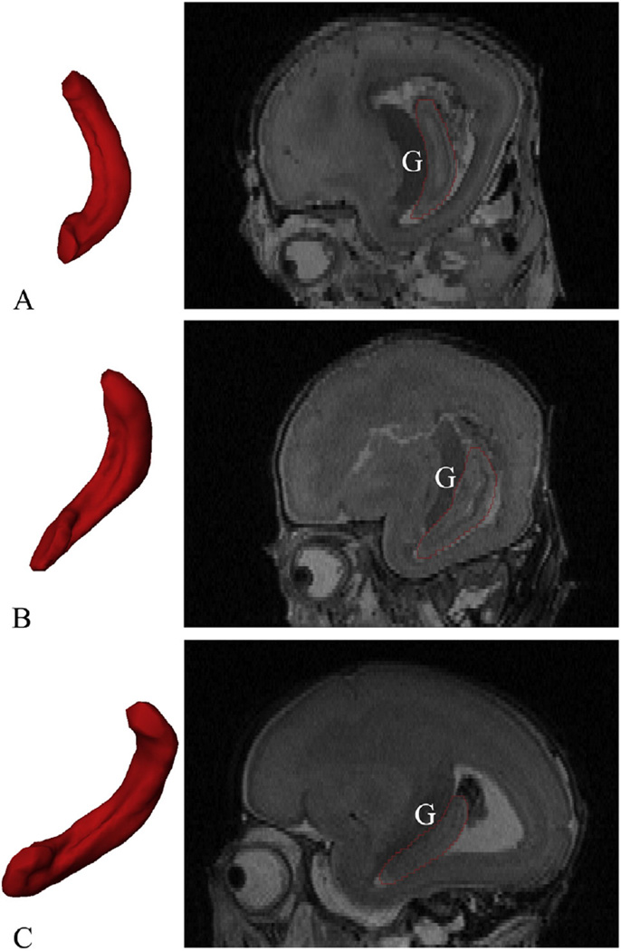
Fig. 7.
Positioning changes of the fetal HF. The left 3Dmodels are the 3D representations of the right HF reconstructed from the segmented masks (medial view, relative volume), and the right sections are the same sagittal section passes through the germinal matrix (G). The whole HF is becoming horizontal as its head folding into the temporal lobe. From top to bottom: (A) 15 GW; (B) 18 GW; and (C) 21 GW.

