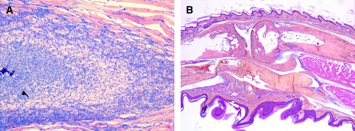
Figure 3.
Histological observation of mouse footpads from (A) group A (exposed to Mycobacterium ulcerans Agy99-inoculated soil) mice and (B) group C mice (negative control). In the group A mice, but not in the group C mice, we observed an abundance of inflammatory infiltrate composed of neutrophils and foamy macrophages (Giemsa stain, original magnification ×100).

