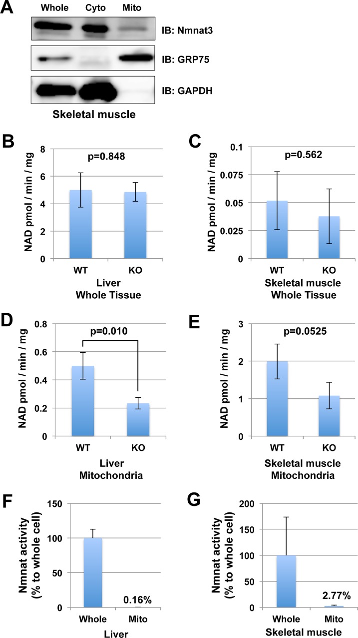
Fig 3. Nmnat3 partially contributes to the Nmnat activity in mitochondria.
(A) Subcellular fractionation of endogenous Nmnat3 protein in skeletal muscle. GAPDH was used as a cytoplasmic fraction marker, and GRP75 (mitochondrial HSP70) was used as a mitochondrial fraction marker. (B and C) Nmnat activities of total tissue lysates from liver (B) and skeletal muscle (C) were measured by enzymatic activity assay. For this assay, tissue lysates were prepared from WT and Nmnat3 KO mice. Samples were dialyzed to remove endogenous metal ion and metabolites. Data are presented as mean ± SD (n = 4 for each group). (D and E) Mitochondrial Nmnat activities from liver (B) and skeletal muscle (C) were also measured in the same way as above. (F and G) Ratio of Nmnat activity in mitochondria against that in whole cell from WT liver (F) and WT skeletal muscle. Data are presented as mean ± SEM (n = 3 for each group).

