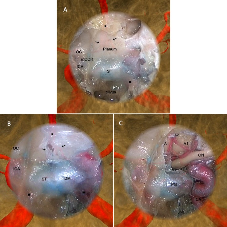
Fig 7. The augmented reality navigation system display during the cadaver head experiment.
After maxillary sinus expansion, ethmoidectomy, and frontal sinus expansion, a wide sphenoidotomy using an endoscopic endonasal approach was performed as the final part of the operation. Moreover, to provide sufficient exposure and maneuverability, the posterior nasal septum and middle turbinate were removed so that the classic intrasphenoid landmarks could be identified (A) involving the planum sphenoidale: OC, optic canal; mOCR, medial optico-carotid recesses; ICA, internal carotid artery; ST, sella turcica; and clivus. In addition, with image superimposition, we could observe the projection of certain anatomic structures on and around the endoscopic image, involving the A1 (black arrows) and A2 (black stars) segments of the anterior cerebral artery and internal carotid artery (black squares). In fact, these structures included any of concern to the surgeon, as long as they were segmented from the computed tomography (CT) image manually or automatically before the operation. (B) Bone in the left superior, posterior, and lateral walls of the sphenoid sinus was removed to expose the dura mater. As the lens moved forward, the projection of the bilateral internal carotid artery became much clearer. (C) After the dura mater was opened, the A1 and A2 segments of the anterior cerebral artery and left internal carotid artery were exposed; the actual locations were consistent with their projections, showing that the virtual and real images were fused accurately and moved synchronously. The AR-N system display expands the surgical field during nasal endoscopy both in terms of depth and breadth, and can thus provide surgeons with more intuitive information regarding anatomical structures.

