
Figure 5.
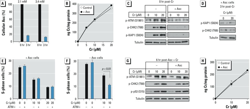
ATM activation in IMR90 normal human lung cells. Control and Asc-restored (+Asc, 3.4 mM Asc) cells were treated with Cr(VI) for 3 hr. Bar and line graphs show means ± SD, n = 3. (A) Asc levels in cells before and after 3-hr incubation in regular medium. (B) Uptake of Cr(VI) by control and Asc-restored (+Asc, 3.4 mM Asc) cells. (C) Western blots of cells collected immediately after Cr(VI) exposure. Images of control and +Asc samples are from the same blots, which were cropped to remove unrelated intervening lanes. (D) Western blots of +Asc cells collected 3 hr after exposure to Cr(VI). (E) Percentage of +Asc cells in S-phase at 24 hr post-exposure. ATM-i (1 μM KU60019) was present during Cr(VI) exposure and subsequent 24-hr incubations. (F) As in panel E, except that cells were treated with Cr(VI) without Asc restoration. (G) ATM activation and p53 phosphorylation in cells treated with Cr(VI) at 6 hr after Asc loading (3-hr Cr exposure, immediate collection). (H) Cr(VI) uptake by cells treated at 6 hr post–Asc loading. In panels G and H, +Asc cells contained 0.110 ± 0.006 mM Asc at the start of Cr exposure.