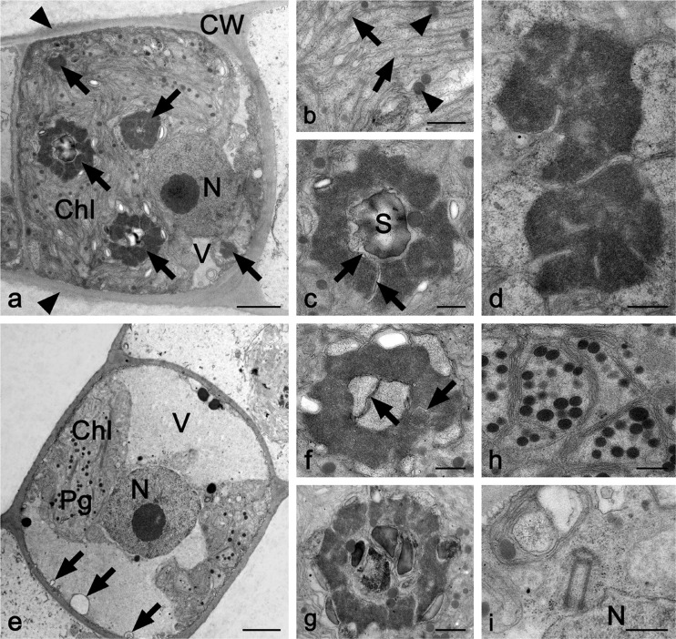
Fig. 2.
TEM micrographs of longitudinal sections through a–d UTEX2353 (Entransia fimbriata) and e–i UTEX2793 (E. fimbriata). a Cell with numerous pyrenoids (arrows), small vacuoles, parietal chloroplast, and nucleus; cell wall covered by thin mucilage layer (arrowheads). b Detail of chloroplast with thylakoid membranes (arrows) and plastoglobules (arrowheads). c Pyrenoid with electron-dense matrix penetrated by thylakoid membranes (arrow) and one large starch grain in the center. d Two pyrenoids. e Cell showing one large vacuole, parietal chloroplast, and central nucleus. Retracted cytoplasm from the cross-wall forms spherical invagination (arrows). f Pyrenoid with thylakoid membranes in the center (arrows) and surrounded by few starch grains. g Pyrenoid with starch grains in the center. h Detail of chloroplast with numerous plastoglobules. i Two centrioles close to the nucleus. Chl chloroplast, CW cell wall, N nucleus, Pg plastoglobules, S starch grain, V vacuole. Bars a, e 2 μm; b–d, f–i 500 nm

