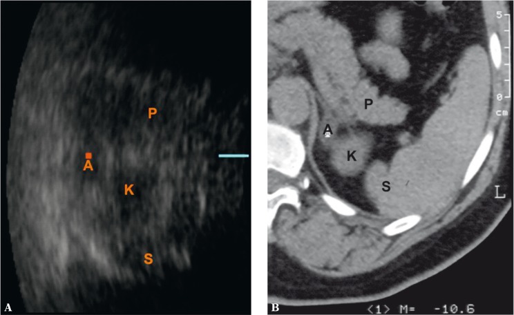
Fig. 3.
Slight adenoma of the left adrenal gland – A. P – pancreas, K – upper renal pole, S – spleen. Three-dimensional ultrasound – axial image (A). This slight tumor of the left adrenal gland was not detected in a real-time 2D examination. 3D ultrasound enables the lesion to be visualized and its size to be compared with CT (B)

