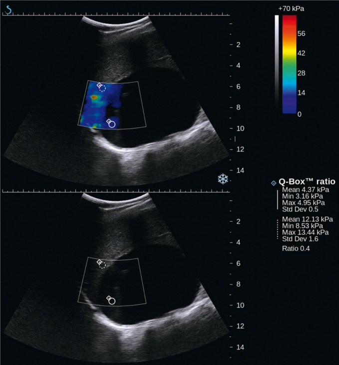
Fig. 4.
Recurring lymphatic cyst of the right adrenal gland of the maximum size of 10 cm: shear-wave elastography (SWE) – upper image, B-mode – lower image. In the upper SWE image, there is no colored signal since shear waves do not propagate through fluids. Inside the lesion at its periphery, an artefact-like colored signal can be seen, however indicating low stiffness values (approximately 4 kPa)

