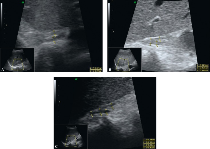
Fig. 7.
Ultrasound B-mode image of the normal right adrenal gland with marked measurements of the thickness of the limbs and body (calipers). A. Axial plane with harmonic imaging. Hypoechoic adrenal gland of V shape. The echogenicity of the adrenal limb ends is similar to the echogenicity of the retroperitoneal fat tissue. B. Longitudinal section through the adrenal gland. In this view, the adrenal gland is of λ shape. It is only slightly hypoechoic in relation to the retroperitoneal fat tissue. C. Longitudinal section with harmonic imaging: increased contrast between the hypoechoic adrenal gland and the retroperitoneal fat tissue (compared with Fig. 7B). Harmonic imaging is recommended in the visualization of normal adrenal glands and their focal lesions

