Fig. 1.

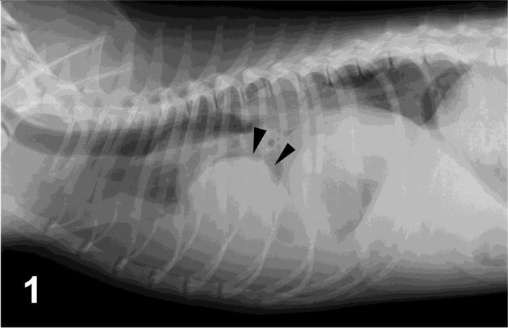
Case No 1. Right lateral thoracic radiograph. There is a large mass in the right middle and caudal lung lobes (arrowheads), obscuring the apex of the cardiac silhouette on the lateral view.

Case No 1. Right lateral thoracic radiograph. There is a large mass in the right middle and caudal lung lobes (arrowheads), obscuring the apex of the cardiac silhouette on the lateral view.