Fig. 2.

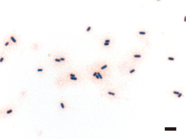
Photomicrograph of a peripheral blood smear sample with Gram stain. Several boxcar-shaped, Gram-positive bacilli can be seen. Bar: 5 µm.

Photomicrograph of a peripheral blood smear sample with Gram stain. Several boxcar-shaped, Gram-positive bacilli can be seen. Bar: 5 µm.