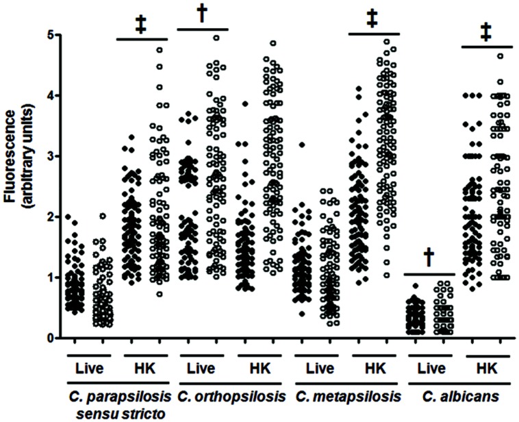
FIGURE 1.
Analysis of β1,3-glucan and chitin exposure at the cell wall surface. Live (L) yeast cells were incubated with IgG Fc-Dectin-1and then with FITC-conjugated IgG to label β1,3-glucan (closed circles). Alternatively, chitin was labeled with WGA-FITC (open circles). The fluorescence associated to each cell is plotted. In addition, the experiment was conducted with cells previously heat-inactivated (HK). Strains used are Candida parapsilosis sensu stricto SZMC 8110, C. orthopsilosis SZMC 1545, C. metapsilosis SZMC 1548, and C. albicans SC5314. One hundred cells were analyzed for each group. ‡P < 0.05, when compared with the live cells; †P < 0.05 when compared to the other groups with live cells.

