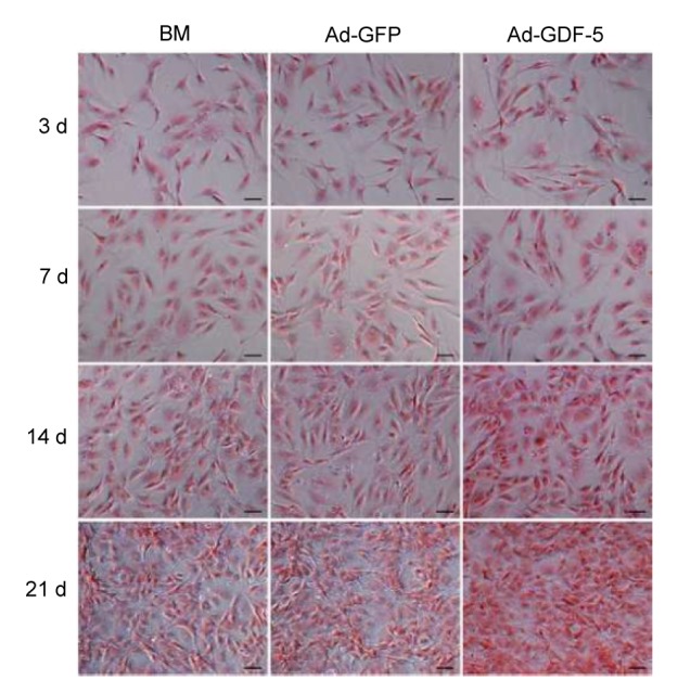
Fig. 5.

Safranin-O staining (red) of the proteoglycan-rich ECM of human
NP cells NP cells were stained at 3, 7, 14, and 21 d after being infected by Ad-GDF-5, Ad-GFP, or no adenovirus (BM group). Scale bar=50 μm (Note: for interpretation of the references to color in this figure legend, the reader is referred to the web version of this article)
