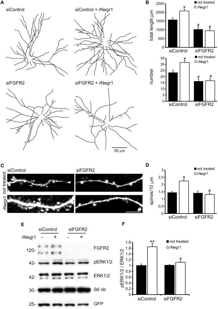
Figure 6.
Negr1 modulates neuron morphology via FGFR2. Cortical neurons were infected at DIV4 with virus expressing siControl or siRNA against FGFR2 (siFGR2) and treated at DIV10 with recombinant Negr1 (40 ng/ml, rNegr1). Neurons were processed for immunofluorescence at DIV18 and infected GFP positive neurons imaged via confocal microscopy. Panels show camera lucida tracing (A). Graphs show neurite total length and number (B). Data are reported as mean ± SEM; *p < 0.001 vs. not treated, same infection, #p < 0.01 vs. siControl, same treatment. Scale bar = 50 μm. Dendritic spines were recognized as mushroom like protrusions decorating the neurites (C). Spine density was calculated as protrusion number along 10 μm of neuritic length. Data are reported as mean ± SEM; *p < 0.001 vs. not treated, same infection, #p < 0.01 vs. siControl, same treatment. Scale bar = 10 μm (D). Cortical neurons were infected with virus expressing siControl or siRNA against FGFR2 (siFGR2) and treated with recombinant Negr1 (40 ng/ml, rNegr1 10 min). Neurons were processed for western-blotting to investigate ERK1/2 phosphorylation (E). The graph reports p-ERK1/2 level normalized vs. total ERK1/2 amount (F). Data are expressed as mean ± SEM n = 5. **p < 0.01 vs. not treated, same infection; #p < 0.01 vs. siControl, same treatment.

