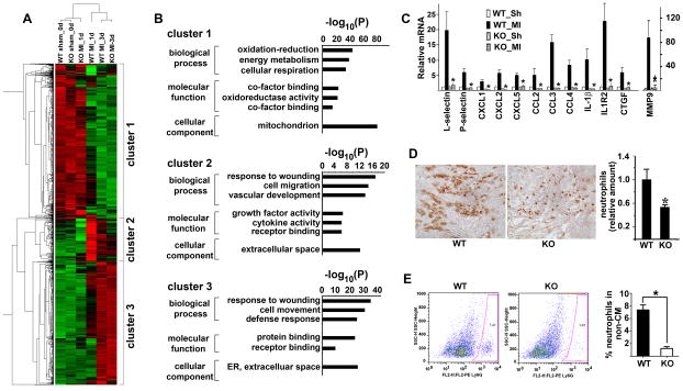
Figure 2. Deletion of FoxO4 resulted in attenuated early post-MI inflammation.
(A) Heat map of differentially expressed genes (>2-fold changes) in WT and FoxO4-nll mouse hearts at sham and post-MI 1 and 3 days. (B) Gene ontology analysis showing the biological processes and molecular functions of differentially expressed genes in (A). (C) qRT-PCR of inflammatory genes that are most attenuated in one-day post-MI FoxO4 KO mice. Expression was normalized against GAPDH and expressed relative to that of WT_Sham (Sh) heart (n=5). *, p<0.05 compared to WT_MI. (D) Representative images of Ly6G staining of sections of WT and FoxO4 KO mouse hearts at one day post-MI. Ly6G staining was quantified by Image-J and expressed relative to the mean value for WT mouse hearts (n=5–7). *, p< 0.05. (E) FACS profiles of non- cardiac mycoytes labeled with Ly6G (n=5–7), *, p<0.05.

