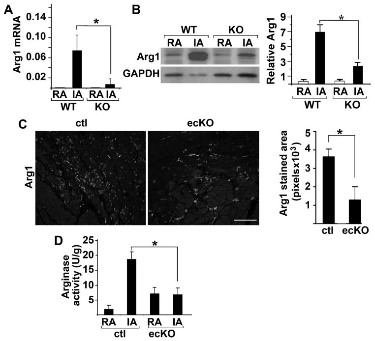
Figure 4. Upregulation of post-MI Arg1 expression was significantly attenuated in FoxO4 KO and FoxO4 ecKO mouse hearts.
(A) Relative mRNA of Arg1 in the remote (RA) and infarct area (IA) of FoxO4 KO and WT littermate mouse hearts post-MI 1 day as measured by qRT-PCR. mRNAs were normalized against internal GAPDH (n=6). *, p<0.05. (B) Western blot of Arg1 from tissues described in (A) (n=3). *, p<0.05. Protein levels were quantified by densitometry and normalized against GAPDH. (C) Immunofluorescent micrographs of IA from histological heart sections of FoxO4 ecKO and control littermates stained with Arg1 antibody post-MI day 2. Arg1 immunofluorescence from 5 different sections of each genotype mouse was quantified by Image-J, and averaged values are shown (n=3) *, p<0.05. Scale bar=100μM. (D) Arginase activity of lysates from remote area (RA) and infarct area (IA) of FoxO4 ecKO or control littermates (n=3). *, p<0.05.

