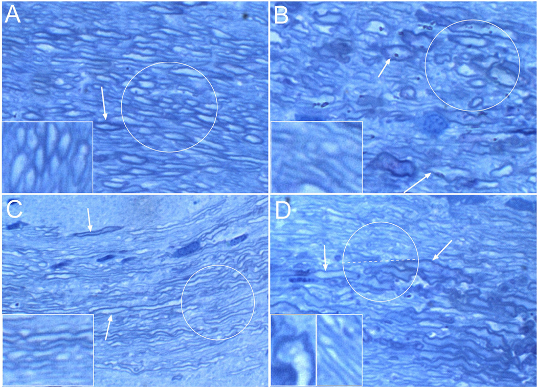
Figure 1. Ethanol and NNK exposures cause structural damage to white matter.
Representative images of corpus callosum white matter from (A) control, (B) NNK-exposed, (C) ethanol-fed, or (D) ethanol+NNK treated Long Evans rats. Brain tissue samples were fixed in glutaraledhyde and embedded in epon. 1-µm thick sections were stained with Toluidine blue. (A) Note the abundant large, well-myelinated fibers (highlighted with the circle and shown at higher magnification in the inset). Arrow points to a thickly myelinated axon. (B) Relative to control, myelinated fibers in NNK-exposed corpora callosa are highly irregular in size and shape due to combined effects of dystrophic enlargement and atrophy. Demyelination marked by reduced intensity of myelin sheath staining (toluidine blue-stained borders; see inset). Upper central arrow shows an enlarge well-myelinated axon, while the lower arrow show an irregularly enlarged partly demyelinated axon. (C) The main effect of chronic ethanol-exposure was demyelination. The majority of axons are thinly myelinated and they appear to be much smaller in caliber than control (circle; also compare inset images). The upper arrow shows some preservation of myelin around one axon, while the lower arrow shows demyelination of a relatively large axon. (D) Ethanol+NNK treatments produced effects corresponding to both NNK and ethanol exposures. Note that in the upper half of the circled fibers and right side of the inset, the fibers mainly show demyelination, whereas in the lower half of the circle, at the arrow tips, and in the left inset, the fibers are mainly dystrophic. Original magnification, 650× (Insets, 1200×).

